This 3D printer can repair damaged tissue from within
This flexible 3D printer works from the inside out to repair tissues and organs, which researchers say is an extremely promising invention.
Researchers at the University of New South Wales (UNSW) in Sydney have developed a flexible 3D bioprinter capable of creating layers of organic materials directly on organs or tissues. Unlike other bioprinting approaches, this system is considered minimally invasive and could in some cases help avoid major surgery or organ removal – at least in theory – but scientists warn that it will be another five to seven years before the first tests on people. completed.
This flexible 3D printer works from within to regenerate tissues and organs.
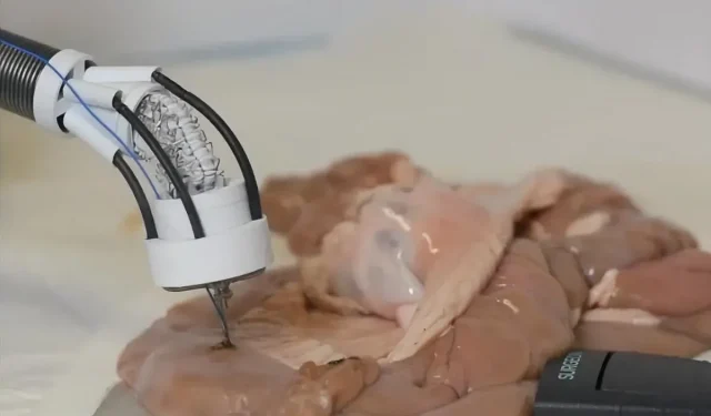
This printer, called the F3DB, has a soft robotic arm capable of harvesting living cell biomaterials on injured internal organs or tissues. Its flexible serpentine body enters the body through the mouth or anus, and the surgeon/driver gestures it towards the area to be repaired. In addition, the robot has small cannons to deliver water to targeted areas, and its print head can also act as an electric scalpel. The team hopes that its multifunctional approach will one day become a versatile tool (cut, clean and impression) for minimally invasive surgeries.
The F3DB robotic arm uses soft bellows actuators, which are a hydraulic system consisting of “syringes driven by an AC motor that delivers water to the actuators,”as IEEE Spectrum summarizes it. Its arm and flexible print head can move with three degrees of freedom, just like modern desktop 3D printers. In addition, the device has a flexible miniature camera that allows the operator to visualize what he is doing in real time.
An extremely promising invention, according to researchers
The research team conducted their first lab tests of a version using non-biological materials, namely chocolate and liquid silicone. They then tested this on a pig kidney before moving on to biomaterial prints on a glass surface in an artificial colon. “We saw the cells grow every day and quadruple in seven days, the last day of the experiment,” said Thanh Nho Do, co-leader of this group and senior lecturer at the Graduate School of Biomedical Engineering at the University of New South Wales. “The results show that the F3DB has great potential to become a versatile endoscopic tool for endoscopic submucosal dissection procedures.”
The team is convinced that this device is very promising, but it still has to go through many tests before it can be used in the real world. The next steps will be to continue animal experiments. Thanh Nho Do estimates that it could take five to seven years, but according to Ihrabim Ozbolat, professor of engineering and mechanics at Pennsylvania State University, “commercialization is only a matter of time.”
Leave a Reply